The primary action is to elevate the mandible and laterally deviate it to the opposite side. Specifically the medial pterygoid muscle functions to.
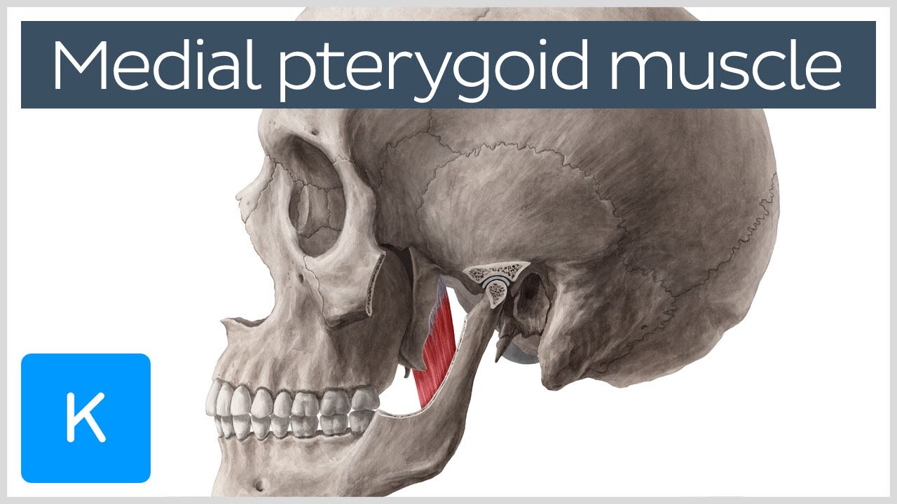
Medial Pterygoid Muscle Origin Insertion Function Nerve Supply Anatomy Kenhub Youtube
Medial pterygoid is a thick quadrilateral muscle that connects the mandible with maxilla sphenoid and palatine bones.
. Method two Though not direct this method helps treat the distal part of. It receives blood supply from the pterygoid branches of the maxillary artery. Actions The medial pterygoid acts together with masseter to elevate the mandible.
The medial pterygoid muscle is a thick and square shaped muscle. Learn about the origin insertion functions and innervation of the medial pter. By contracting on one side the medial pterygoid pushes the mandible to the opposite side.
It has two heads of origin. Medial pterygoid muscle View Related Images. The muscle is right there.
The bilateral contraction of this muscle elevates the mandible and closes the mouth. The deep head is the major component and is attached to the medial aspect of the lateral pterygoid plate of the sphenoid bone. Attachments of Medial Pterygoid Muscle.
From tuberosity of maxilla adjoining boneDeep head. The medial pterygoid muscle attaches to the angle of the mandible and to the lateral pterygoid plate to form a sling with the masseter muscle that suspends the mandible Figure 6-19. Apply mild to moderate pressure and wait for it to relax.
Hold until it relaxes. It can assist in protrusion of the mandible. It belongs to the group of masticatory muscles along with the lateral pterygoid masseter and temporal muscles.
Medial surface of lateral pterygoid plate and palatine bone. Where are the Pterygoids. The action of the muscle during bilateral contraction of the entire muscle is to elevate the mandible raising the lower jaw.
Medial surface of ramus and angle of mandible. It arises from the medial surface of the lateral pterygoid plate and the grooved surface of the pyramidal process of the palatine bone. The medial pterygoid muscle is innervated by the medial pterygoid branch of the mandibular division of the trigeminal nerve CN V3.
Elevation of the mandible occurs during the closing of the jaws. From medial surface of lateral pterygoid plate adjoining pro. The smaller superior head arises from the infratemporal crest of the greater sphenoid bones higher wing while the larger inferior head emerges from the lateral surface of the lateral pterygoid plate.
The superficial head is attached to the maxillary tuberosity and the pyramidal process of the palatine bone. Acting together with the lateral pterygoid muscles they protrude the mandible which is important in the grinding movement of mastication. Raiseelevate the lower jaw.
The origin extends from the superior margin of the pterygoid plate and skirts medially of foramen spinosum and ovale finally terminating on the carotid canal rim. Perform once 1 to 2 times a day. This muscle also divides into two parts.
It is also known as internal pterygoid muscle. The exact origin of the superficial head of the Medial Pterygoid is Maxillary tuberosity and pyramidal process of bone kind of where your wisdom teeth are. Blood supply The arterial supply to medial pterygoid muscle is from the pterygoid branches of the maxillary artery.
The medial pterygoid muscle is one of the four paired muscles of mastication. Although having different origins both heads insert on the inner. It can assist in protrusion of the mandible.
Posterior and inferior border of the mandibular ramus and angle from the mandibular foramen to the mylohyoid groove Actions. But cases involving the medial pterygoid temporal sternocleidomastoid mental platysma scalenus omohyoid buccinator and genioglossus muscles have also been reported. Medial surface of the lateral plate of the pterygoid process pyramidal process of the palatine bone and the maxillary tuberosity Insertion.
What is the action of the masseter muscle. The medial pterygoid muscle a major elevator of the jaw is a square-shaped masticatory muscle located on the medial aspect of the lower jaw bilaterally. The medial pterygoid muscle attaches to the angle of the mandible and to the lateral pterygoid plate to form a sling with the masseter muscle that suspends the mandible Figure 6-19.
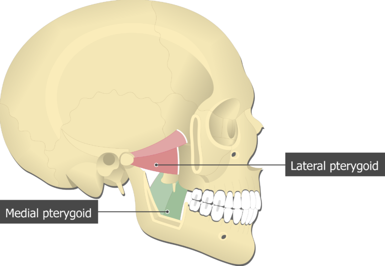
Medial Pterygoid OriginSuperficial head. Koritzer RT Suarez F. The pterygoid muscles are the two jaw muscles of mastication located in the head on the inner surface of the mandible.
This muscle lies medial to the lateral pterygoid muscle. Structure and Function The medial pterygoid muscle has a triple function. The medial pterygoid is a thick quadrilateral muscle.
It has a second slip of origin from the lateral surfaces of the pyramidal process of the palatine and tuberosity of the maxilla. Action Upon bilateral contractions the medial pterygoid muscles push the mandible forward protrude the mandible. Use your time efficiently and maximize your retention of key facts and definitions with study sets created by other students studying Medial Pterygoid Muscle Action.
A previously undescribed accessory medial pterygoid muscle occurred uniformly in the dissection room population studied. Look it up now. In contrast the lateral pterygoid muscle is rarely affected with only one case of isolated unilateral MOT reported in the literature probably caused by a severe form of.
Medial pterygoid muscle consists of two heads. The primary action is to elevate the mandible and laterally deviate it to the opposite side. Raiseelevate the lower jaw Protrude the lower jaw Move the lower jaw side-to-side These movements of the.
Lateral Medial Pterygoid Muscle The pterygoid muscles are two of the four muscles of mastication located in the infratemporal foss a of the skull. The contraction of the medial pterygoid elevates the mandible jaw closure and moves it forward protrusion. Portable and easy to use Medial Pterygoid Muscle Action study sets help you review the information and examples you need to succeed in the time you have available.
Function The medial pterygoid muscle has functions including elevating the mandible closing the mouth protruding the mandible mastication especially for when the maxillary teeth and the mandibular teeth are close together and excursing the mandible contralateral excursion occurs with unilateral contraction. Accessory medial pterygoid muscle. Specifically the medial pterygoid muscle functions to.
Since the medial pterygoid muscle attaches to the lower jaw the function of this muscle is to move the lower jaw. This muscle also contributes to the elevation of the mandible acting as a synergist to the temporalis and masseter. The action of the Temporal Muscle is the elevation and retraction of the mandible which closes the mouth Saladin 2020 p.
Open your jaw and slide your finger in to the inner side of that bone.
![]()
Medial Pterygoid Origin Insertion Action Innervation Kenhub

Medial Pterygoid Muscle Wikipedia

The Muscular System
![]()
Lateral Pterygoid Muscle Medial Pterygoid Muscle Temporal Muscle Masseter Muscle Pterygoid Processes Of The Sphenoid Others Angle Head Anatomy Png Pngwing
Static And Functional Anatomy Of The Human Masticatory System Pocket Dentistry

Medial Pterygoid Muscle Attachments Actions Innervation
Medial Pterygoid Muscle Attachments Actions Innervation
![]()
Medial And Lateral Pterygoid Muscle Anatomy And Function Kenhub
0 comments
Post a Comment